
Fri, Oct 3, 2025

Volume 18, Issue 1 (Jan-Feb 2024)
mljgoums 2024, 18(1): 12-15 |
Back to browse issues page


Download citation:
BibTeX | RIS | EndNote | Medlars | ProCite | Reference Manager | RefWorks
Send citation to:


BibTeX | RIS | EndNote | Medlars | ProCite | Reference Manager | RefWorks
Send citation to:
Shirkhani S, barari A, Abbasi Daloii A, saravi M. Evaluating the expression of NOs and NOX2 genes in patients with coronary artery occlusion following aerobic exercise and Omega-3 intake. mljgoums 2024; 18 (1) :12-15
URL: http://mlj.goums.ac.ir/article-1-1332-en.html
URL: http://mlj.goums.ac.ir/article-1-1332-en.html


1- Department of Sport Physiology, Ayatollah Amoli Branch, Islamic Azad University, Amol, Iran
2- Department of Sport Physiology, Ayatollah Amoli Branch, Islamic Azad University, Amol, Iran ,alireza54.barari@gmail.com
3- Department of Cardiology, Babol University of Medical Sciences, Babol, Iran
2- Department of Sport Physiology, Ayatollah Amoli Branch, Islamic Azad University, Amol, Iran ,
3- Department of Cardiology, Babol University of Medical Sciences, Babol, Iran
Full-Text [PDF 420 kb]
(870 Downloads)
| Abstract (HTML) (2906 Views)
Full-Text: (697 Views)
Introduction
Cardiovascular diseases, especially coronary artery problems, are the main causes of death (1). With increasing urbanization in the developing world, the prevalence of cardiovascular risk factors is observed worldwide and will probably become the most common cause of death worldwide by 2020 (2). Decreased elasticity of large arteries and impaired vascular endothelium are two important factors affecting vascular function that occur with age. Endothelial dysfunction seems to be an important factor in the development of atherosclerosis, hypertension, and disorders (3). Some research has shown that nitric oxide (NO) levels decrease with age. In their study, Topraki et al. showed that the greatest reduction in NO was observed between the ages of 46 and 60 years (4). Recently, NO has been considered an important intermediate in a variety of physiological functions such as nerve conduction, blood pressure regulation, vasodilator, and immune and defense activity. It plays a functional role in all stages of inflammation, and muscles regulate Ca2 dynamics under the influence of NO concentration (5). This gas also relaxes the arteries and airways in the respiratory system, and angiogenesis, apoptosis, cell cycle, invasion, and metastasis are also affected by NO secretion and concentration. It acts as a messenger molecule and exerts its effects through the production of cyclic guanosine monophosphate cGMP. In the body, NO is produced by the enzyme NO synthase from the amino acid al-arginine. In cardiac patients, the reduction of NOX2 prevents oxidative stress and progression of heart disease (6). On the other hand, physical exercises can reduce the risk of coronary artery disease by increasing the maximum oxygen consumption and favorable hemostatic effects. The antithrombotic effects of physical exercise include increased plasma volume, decreased viscosity, decreased platelet adhesion, and increased thrombolytic ability. Intense physical exercise increases fibrinolytic activity by increasing the endothelial synthesis of tissue-activating factor plasminogen-1 (7). Exercise increases mechanical blood flow and causes mechanical stimulation in the arteries, and if the endothelium is healthy, it leads to increased production and release of NO (3). The beneficial effects of endurance exercise in the prevention and reduction of cardiovascular disease have been shown in many studies. In some studies, exercise has shown significant improvements in cardiovascular function and quality of life in patients with heart failure (8). Moderate to long-term physical exercise may improve vascular endothelial function, reduce environmental resistance, and alter autonomic function, all of which may improve functional capacity (9). Numerous studies have shown that aerobic exercise reduces arterial stiffness in healthy individuals of all ages, aerobic competition champions, and coronary artery disease patients (10). Therefore, many researchers have concluded that the implementation of exercise programs by patients improves the performance of physical and mental fitness, reduces the risk of heart attack, reduces heart rate and systolic blood pressure, and increases the amount of oxygen consumption of resting heart muscle. Increase in capacity of aerobic activity related to reduces anxiety and depression and increases in optimism. In addition, researchers and cardiovascular specialists consider regular exercise to be beneficial and safe for most patients after myocardial ischemia (11). Despite being new in Iran, cardiac rehabilitation has found special importance and place in the treatment of patients with coronary heart disease. These exercises increase the efficiency of oxygen extraction and skeletal muscle metabolism, reduce heart function, and increase coronary blood flow. In addition, epidemiological studies have shown that regular exercise can reduce cardiovascular mortality in people with a history of heart disease due to its physiological effects. Exercise has a beneficial effect on cardiovascular adaptation, which can vary depending on the type, intensity, and duration of exercise (12). People with higher levels of physical activity are less likely to die from coronary artery disease (12,13). Exercise seems to be not only a tool to maintain a healthy lifestyle but also a safe prescription for prevention (14). The aim of the research in this direction is to evaluate the expression of NO synthase (NOS) and nicotinamide adenine dinucleotide oxidase (NOX2) genes in patients with coronary artery occlusion following aerobic exercise and omega-3 intake.
Methods
The design of the present study was quasi-experimental, with a pre-test and post-test. The statistical population in this study was all men with cardiovascular diseases in the age range of 55 to 65 years visiting Rouhani and Shahid Beheshti hospitals in Babol, Iran (in the second half of 1398) without a history of regular exercise and without a history of omega-3 consumption (They had not taken omega-3 supplements for at least the past 6 months) who were selected by responding to a call. From all interested patients, 32 were selected as the sample. These people participated in this study after medical examinations, completing a questionnaire, and providing consent. These individuals were randomly divided into 4 groups: control, omega-3, exercise, and omega-3 + exercise. This study was approved by the Ethics Committee at the Islamic Azad University, Babol Branch (reference number: IR.IAU.ABOL.REC.1398.092).
The training program consisted of 8 weeks of intermittent running training, 3 sessions per week, with an intensity of 55 to 65% of the subjects' heart rate reserve (HRR) and with an emphasis on gradual overload. The training protocol was to run indoors. In these exercises, the beginning of each exercise session began with 10 minutes of general warm-up, including stretching, light and dynamic movements of the whole body, and, at the end, 10 minutes of cooling. The main workout that was set to reach intensity was monitored using a polar clock to show heart rate (15).
"The participants took a daily dose of 1000 mg capsules each morning (containing EPA 180 and DHA 120) from the Viva Omega-3 fish oil brand manufactured in Canada (16,17). It is better to take an omega-3 supplement with a meal because it is a kind of oil supplement and is better absorbed with food.
Blood samples were collected from the subjects 24 hours before and 48 hours after the last training session after a night of fasting, and the serum was separated by centrifugation.
To extract RNA, about 100 μL of the Buffy Coat was placed in a microtubule free of RNase enzyme, and 1 cc of TRIzol solution was added. The microtubes were centrifuged at 2-8 °C for 15 minutes at 12000 g (18,19). Finally, the precipitate was dissolved in DEPC-treated water and stored in a freezer at -70 ° C. Except for the first stage, which was performed under a conventional hood due to TRIzol toxicity, all the steps were performed under a laminar hood. The extracted RNA was quantitatively analyzed by spectrophotometry and electrophoresis on agarose gel. The cDNA was fabricated based on the Fermentas kit (20). The primers were designed using Gene Runner version 6.0 and Oligoanalyzer version 1.0 software for oligonucleotides, enabling the analysis and design of DNA oligos to understand their properties and behavior. After designing, their specificity was verified using BLAST software on the NCBI (National Center for Biotechnology Information) online platform to ensure complete specificity and uniqueness to the desired genes. Table 1 shows the 2 designed primers.
After reverse transcription reaction, real-time polymerase chain reaction (PCR) was performed using the SYBR Green method to amplify the desired fragment and quantitatively evaluate the expression of genes on the cDNA (21). The LinRegPCR software version 1.0 was used to determine the efficiency of primers. In this software, a group is determined for the samples working with a pair of primers, and for each group (each pair of primers), an efficiency is obtained. After examining all real-time PCRs, the efficiencies obtained from all of them were averaged, and final efficiencies were determined. After performing the real-time PCR and collecting raw data, they were reviewed and analyzed.
Statistical data analysis
Descriptive statistics was used to describe the data. Within-group and between-group changes from the pre-test to the post-test were examined by 2-way analysis of variance with repeated measures and, then, Tukey's test. The significance level in all cases was α<0.05. All statistical tests were performed using Graph Pad Prism 8 and Microsoft Excel 16 software at a significant level of P <0.05
Results
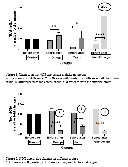
The results of Table 2 indicate a significant difference in the expression of NOS and NOX genes between different groups from pre-test to post-test (P <0.0001). The results of within-group changes showed that the mean expression of the NOS gene in the exercise groups (P = 0.0224) and the combination of exercise + omega (P <0.0001) increased significantly from pre-test to post-test. However, in the omega group, there was a nonsignificant increase (P = 0.5667). The results of Tukey's post-hoc test also showed that there was a significant increase (P <0.0001) between the mean expression of the NOS gene in the exercise + omega group compared to the control group. However, the exercise group (P ≥ 0.9999) and omega group (P = 0.8519) had a significant increase compared to the control group. The omega group was not significantly different from the exercise group (P = 0.9530). The omega + exercise group had a greater and significant increase (P <0.0001) than the omega and exercise groups (Figure 1). Also, the results of within-group changes showed that the mean ratio of NOX gene expression changes in the exercise, omega, and combination of exercise + omega groups from pre-test to post-test had a significant decrease (P <0.0001). The results of Tukey's post-hoc test also showed that the mean ratio of NOX gene expression changes in the exercise, omega, and combination of exercise + omega groups compared to the control group had a significant decrease (P <0.0001). Nevertheless, there was no significant difference between exercise + omega, exercise, and omega groups. However, the omega + exercise group showed a further decrease (Figure 2).
Discussion
The results of the present study showed that the mean expression of the NOS gene in the exercise groups and a combination of exercise + omega from pre-test to post-test increased significantly, and in the omega group, it had a nonsignificant increase. The omega + exercise group had a greater and more significant increase than the omega and exercise groups. Also, the mean ratio of NOX gene expression changes in training groups, omega, and combination of training + omega from pre-test to post-test had a significant decrease. However, the omega + exercise group showed a further decrease. NADPH oxidases (NOX) are enzymatic assemblies that have been considered key molecules in vascular dysfunction. NOX has the main function of producing reactive oxygen species (ROS) and is the main source of ROS production in endothelial cells (22). The endothelium is a thin layer that covers the inner surface of blood vessels and acts as a secretory organ to maintain blood flow homeostasis. Enzymatic production of NO by endothelial NO synthase (eNOS) is very important in mediating endothelial function, and oxidative stress can lead to improper regulation of eNOS and endothelial dysfunction (23). However, cardiovascular disease 2 is characterized by poor control of the endothelial cell redox environment by a change in the direction of overproduction of ROS by NOX (24). In heart patients, reducing NOX2 prevents oxidative stress and the progression of heart disease (6). The results of this study show one of the most important roles of NOX2 pathogen in heart failure and strengthening the human heart. Obesity and eating saturated fats increase the risk of heart failure and arrhythmias. The physiological level of saturated fat can increase mitochondrial ROS in cardiomyocytes and lead to abnormalities in calcium homeostasis and mitochondrial function. PKC or NOX2 inhibitors prevent dysfunction and increase mitochondrial ROS (25). In adult cardiomyocytes, palmitate has been shown to induce ROS by a mechanism that requires mitochondrial uptake and beta-oxidation of palmitate, and mitochondrial ROS is enhanced by PKC-NOX2 activation. Activation of this pathway leads to an increase in sarcoplasm. Reticulum calcium leakage leads to mitochondrial calcium overload and mitochondrial dysfunction. Drug inhibition of NOX2 prevents abnormalities caused by saturated fat (25). On the other hand, NO acts as a messenger molecule and exerts its effects through the production of cyclic guanosine monophosphate cGMP. The role of omega-3 in reducing oxidative damage and restoring free radical homeostasis is not fully understood. Although some studies suggest that omega-3 components may reduce oxidative damage in humans and animals, the data are still inconclusive. There is evidence that omega-3s at least improve the status of antioxidant enzymes in humans. In elderly patients who are chronically exposed to particulate matter, fish oil (2 g capsule per day; 52.4% DHA, 25.0% EPA, and 5.8% DPA) improves antioxidant status (26). Interventional studies confirmed that the use of n-3PUFA provides benefits for the primary and secondary prevention of cardiovascular disease. Evidence from cellular and molecular research studies suggests that the protective effects of n-3 PUFA on the heart are due to synergies between complex and multiple mechanisms, including anti-inflammatory, lipid-mediated dissolution, modulation of cardiac ion channels, and reduction of triglycerides. This index influences cellular signaling pathways, as well as antithrombotic and antiarrhythmic effects. The n-3 PUFAs inhibit inflammatory signaling pathways (27). Activated oxygen and nitrogen species regulate a wide range of signaling pathways that govern cardiovascular physiology. However, oxidant stress due to impaired oxidation signaling has adverse effects on the pathogenesis and progression of cardiovascular disease (28). In addition to increasing eNOS and improving myocardial protection, voluntary physical activity has been shown to alter the expression and phosphorylation of eNOS according to the type of tissue and isomerism of the enzyme - eNOS. These findings indicate that there is a significant difference between exercise-induced myocardial protection and other types of delayed preparation, such as heat pressure and ischemic preparation. The endothelium plays an important role in the protective effects of exercise, especially as increased shear stress on the vessel wall due to exercise increases the expression and activity of vascular eNOS, thereby increasing the production and bioavailability of NO throughout the body. In this study, it was found that intermittent exercise was associated with increased NOS expression (29). Due to the role of inflammation in the pathogenesis of cardiovascular disease, one of the mechanisms for increasing NOS and decreasing NOX in cardiovascular patients may be reducing inflammatory markers through exercise (30). In contrast, skeletal muscle contraction produces ROS and RNS (reactive nitrogen species), which are needed to regulate many of the proteins involved in the stimulation-contraction pair. The amount and species of ROS/RNS produced by contractile muscles will have downstream effects on specific protein targets and cellular oxidation signaling. Redox modifications on specific proteins are essential for the adaptive response to exercise, and skeletal muscle can lead to an unregulated redox reaction during aging (31,32). Exercise provides protective effects against pathological hypertrophy of the heart. Nitric oxide plays an important role in modulating cardiac hypertrophy. However, few studies have examined the relationship between NO signaling and the inhibitory effect of endurance training (ET) on the pathological nature of heart regeneration (33,34).
Conclusion
The results of the present study showed an increase in NOS gene expression and a decrease in NOX gene expression after the simultaneous effect of intermittent exercise and omega-3 supplementation. The ability of exercise and omega-3 supplementation to reduce the level of oxidant stress by-products and increase the control of homeostasis in coronary artery insufficiency suggests an important molecular mechanism underlying the benefits of these interventions.
Acknowledgement
The authors of this article would like to thank the subjects for their cooperation.
Funding sources
The authors of this article did not receive any financial support for the research, writing or publication of this article.
Ethical statement
This study has been approved by the Ethics Committee of the Islamic Azad University, Babol Branch (reference number: IR.IAU.ABOL.REC.1398.092).
Conflicts of interest
All the authors declare that there is no conflict of interest.
Author contributions
This research was carried out under the supervision of Dr. Alireza Barari. The research topic of the original article was finalized by Sedigheh Shirkhani. The obtained data were analyzed and interpreted by Dr. Asieh Abbassi Daloii, and the draft of the manuscript was prepared in collaboration with Dr. Mehrdad Saravi.
Cardiovascular diseases, especially coronary artery problems, are the main causes of death (1). With increasing urbanization in the developing world, the prevalence of cardiovascular risk factors is observed worldwide and will probably become the most common cause of death worldwide by 2020 (2). Decreased elasticity of large arteries and impaired vascular endothelium are two important factors affecting vascular function that occur with age. Endothelial dysfunction seems to be an important factor in the development of atherosclerosis, hypertension, and disorders (3). Some research has shown that nitric oxide (NO) levels decrease with age. In their study, Topraki et al. showed that the greatest reduction in NO was observed between the ages of 46 and 60 years (4). Recently, NO has been considered an important intermediate in a variety of physiological functions such as nerve conduction, blood pressure regulation, vasodilator, and immune and defense activity. It plays a functional role in all stages of inflammation, and muscles regulate Ca2 dynamics under the influence of NO concentration (5). This gas also relaxes the arteries and airways in the respiratory system, and angiogenesis, apoptosis, cell cycle, invasion, and metastasis are also affected by NO secretion and concentration. It acts as a messenger molecule and exerts its effects through the production of cyclic guanosine monophosphate cGMP. In the body, NO is produced by the enzyme NO synthase from the amino acid al-arginine. In cardiac patients, the reduction of NOX2 prevents oxidative stress and progression of heart disease (6). On the other hand, physical exercises can reduce the risk of coronary artery disease by increasing the maximum oxygen consumption and favorable hemostatic effects. The antithrombotic effects of physical exercise include increased plasma volume, decreased viscosity, decreased platelet adhesion, and increased thrombolytic ability. Intense physical exercise increases fibrinolytic activity by increasing the endothelial synthesis of tissue-activating factor plasminogen-1 (7). Exercise increases mechanical blood flow and causes mechanical stimulation in the arteries, and if the endothelium is healthy, it leads to increased production and release of NO (3). The beneficial effects of endurance exercise in the prevention and reduction of cardiovascular disease have been shown in many studies. In some studies, exercise has shown significant improvements in cardiovascular function and quality of life in patients with heart failure (8). Moderate to long-term physical exercise may improve vascular endothelial function, reduce environmental resistance, and alter autonomic function, all of which may improve functional capacity (9). Numerous studies have shown that aerobic exercise reduces arterial stiffness in healthy individuals of all ages, aerobic competition champions, and coronary artery disease patients (10). Therefore, many researchers have concluded that the implementation of exercise programs by patients improves the performance of physical and mental fitness, reduces the risk of heart attack, reduces heart rate and systolic blood pressure, and increases the amount of oxygen consumption of resting heart muscle. Increase in capacity of aerobic activity related to reduces anxiety and depression and increases in optimism. In addition, researchers and cardiovascular specialists consider regular exercise to be beneficial and safe for most patients after myocardial ischemia (11). Despite being new in Iran, cardiac rehabilitation has found special importance and place in the treatment of patients with coronary heart disease. These exercises increase the efficiency of oxygen extraction and skeletal muscle metabolism, reduce heart function, and increase coronary blood flow. In addition, epidemiological studies have shown that regular exercise can reduce cardiovascular mortality in people with a history of heart disease due to its physiological effects. Exercise has a beneficial effect on cardiovascular adaptation, which can vary depending on the type, intensity, and duration of exercise (12). People with higher levels of physical activity are less likely to die from coronary artery disease (12,13). Exercise seems to be not only a tool to maintain a healthy lifestyle but also a safe prescription for prevention (14). The aim of the research in this direction is to evaluate the expression of NO synthase (NOS) and nicotinamide adenine dinucleotide oxidase (NOX2) genes in patients with coronary artery occlusion following aerobic exercise and omega-3 intake.
Methods
The design of the present study was quasi-experimental, with a pre-test and post-test. The statistical population in this study was all men with cardiovascular diseases in the age range of 55 to 65 years visiting Rouhani and Shahid Beheshti hospitals in Babol, Iran (in the second half of 1398) without a history of regular exercise and without a history of omega-3 consumption (They had not taken omega-3 supplements for at least the past 6 months) who were selected by responding to a call. From all interested patients, 32 were selected as the sample. These people participated in this study after medical examinations, completing a questionnaire, and providing consent. These individuals were randomly divided into 4 groups: control, omega-3, exercise, and omega-3 + exercise. This study was approved by the Ethics Committee at the Islamic Azad University, Babol Branch (reference number: IR.IAU.ABOL.REC.1398.092).
The training program consisted of 8 weeks of intermittent running training, 3 sessions per week, with an intensity of 55 to 65% of the subjects' heart rate reserve (HRR) and with an emphasis on gradual overload. The training protocol was to run indoors. In these exercises, the beginning of each exercise session began with 10 minutes of general warm-up, including stretching, light and dynamic movements of the whole body, and, at the end, 10 minutes of cooling. The main workout that was set to reach intensity was monitored using a polar clock to show heart rate (15).
"The participants took a daily dose of 1000 mg capsules each morning (containing EPA 180 and DHA 120) from the Viva Omega-3 fish oil brand manufactured in Canada (16,17). It is better to take an omega-3 supplement with a meal because it is a kind of oil supplement and is better absorbed with food.
Blood samples were collected from the subjects 24 hours before and 48 hours after the last training session after a night of fasting, and the serum was separated by centrifugation.
To extract RNA, about 100 μL of the Buffy Coat was placed in a microtubule free of RNase enzyme, and 1 cc of TRIzol solution was added. The microtubes were centrifuged at 2-8 °C for 15 minutes at 12000 g (18,19). Finally, the precipitate was dissolved in DEPC-treated water and stored in a freezer at -70 ° C. Except for the first stage, which was performed under a conventional hood due to TRIzol toxicity, all the steps were performed under a laminar hood. The extracted RNA was quantitatively analyzed by spectrophotometry and electrophoresis on agarose gel. The cDNA was fabricated based on the Fermentas kit (20). The primers were designed using Gene Runner version 6.0 and Oligoanalyzer version 1.0 software for oligonucleotides, enabling the analysis and design of DNA oligos to understand their properties and behavior. After designing, their specificity was verified using BLAST software on the NCBI (National Center for Biotechnology Information) online platform to ensure complete specificity and uniqueness to the desired genes. Table 1 shows the 2 designed primers.
|
Table 1. Primers used
 |
Statistical data analysis
Descriptive statistics was used to describe the data. Within-group and between-group changes from the pre-test to the post-test were examined by 2-way analysis of variance with repeated measures and, then, Tukey's test. The significance level in all cases was α<0.05. All statistical tests were performed using Graph Pad Prism 8 and Microsoft Excel 16 software at a significant level of P <0.05
Results
The results of Table 2 indicate a significant difference in the expression of NOS and NOX genes between different groups from pre-test to post-test (P <0.0001). The results of within-group changes showed that the mean expression of the NOS gene in the exercise groups (P = 0.0224) and the combination of exercise + omega (P <0.0001) increased significantly from pre-test to post-test. However, in the omega group, there was a nonsignificant increase (P = 0.5667). The results of Tukey's post-hoc test also showed that there was a significant increase (P <0.0001) between the mean expression of the NOS gene in the exercise + omega group compared to the control group. However, the exercise group (P ≥ 0.9999) and omega group (P = 0.8519) had a significant increase compared to the control group. The omega group was not significantly different from the exercise group (P = 0.9530). The omega + exercise group had a greater and significant increase (P <0.0001) than the omega and exercise groups (Figure 1). Also, the results of within-group changes showed that the mean ratio of NOX gene expression changes in the exercise, omega, and combination of exercise + omega groups from pre-test to post-test had a significant decrease (P <0.0001). The results of Tukey's post-hoc test also showed that the mean ratio of NOX gene expression changes in the exercise, omega, and combination of exercise + omega groups compared to the control group had a significant decrease (P <0.0001). Nevertheless, there was no significant difference between exercise + omega, exercise, and omega groups. However, the omega + exercise group showed a further decrease (Figure 2).
|
Table 2. Results of analysis of variance for the ratio of expression changes of NOS and NOX genes
 |
Discussion
The results of the present study showed that the mean expression of the NOS gene in the exercise groups and a combination of exercise + omega from pre-test to post-test increased significantly, and in the omega group, it had a nonsignificant increase. The omega + exercise group had a greater and more significant increase than the omega and exercise groups. Also, the mean ratio of NOX gene expression changes in training groups, omega, and combination of training + omega from pre-test to post-test had a significant decrease. However, the omega + exercise group showed a further decrease. NADPH oxidases (NOX) are enzymatic assemblies that have been considered key molecules in vascular dysfunction. NOX has the main function of producing reactive oxygen species (ROS) and is the main source of ROS production in endothelial cells (22). The endothelium is a thin layer that covers the inner surface of blood vessels and acts as a secretory organ to maintain blood flow homeostasis. Enzymatic production of NO by endothelial NO synthase (eNOS) is very important in mediating endothelial function, and oxidative stress can lead to improper regulation of eNOS and endothelial dysfunction (23). However, cardiovascular disease 2 is characterized by poor control of the endothelial cell redox environment by a change in the direction of overproduction of ROS by NOX (24). In heart patients, reducing NOX2 prevents oxidative stress and the progression of heart disease (6). The results of this study show one of the most important roles of NOX2 pathogen in heart failure and strengthening the human heart. Obesity and eating saturated fats increase the risk of heart failure and arrhythmias. The physiological level of saturated fat can increase mitochondrial ROS in cardiomyocytes and lead to abnormalities in calcium homeostasis and mitochondrial function. PKC or NOX2 inhibitors prevent dysfunction and increase mitochondrial ROS (25). In adult cardiomyocytes, palmitate has been shown to induce ROS by a mechanism that requires mitochondrial uptake and beta-oxidation of palmitate, and mitochondrial ROS is enhanced by PKC-NOX2 activation. Activation of this pathway leads to an increase in sarcoplasm. Reticulum calcium leakage leads to mitochondrial calcium overload and mitochondrial dysfunction. Drug inhibition of NOX2 prevents abnormalities caused by saturated fat (25). On the other hand, NO acts as a messenger molecule and exerts its effects through the production of cyclic guanosine monophosphate cGMP. The role of omega-3 in reducing oxidative damage and restoring free radical homeostasis is not fully understood. Although some studies suggest that omega-3 components may reduce oxidative damage in humans and animals, the data are still inconclusive. There is evidence that omega-3s at least improve the status of antioxidant enzymes in humans. In elderly patients who are chronically exposed to particulate matter, fish oil (2 g capsule per day; 52.4% DHA, 25.0% EPA, and 5.8% DPA) improves antioxidant status (26). Interventional studies confirmed that the use of n-3PUFA provides benefits for the primary and secondary prevention of cardiovascular disease. Evidence from cellular and molecular research studies suggests that the protective effects of n-3 PUFA on the heart are due to synergies between complex and multiple mechanisms, including anti-inflammatory, lipid-mediated dissolution, modulation of cardiac ion channels, and reduction of triglycerides. This index influences cellular signaling pathways, as well as antithrombotic and antiarrhythmic effects. The n-3 PUFAs inhibit inflammatory signaling pathways (27). Activated oxygen and nitrogen species regulate a wide range of signaling pathways that govern cardiovascular physiology. However, oxidant stress due to impaired oxidation signaling has adverse effects on the pathogenesis and progression of cardiovascular disease (28). In addition to increasing eNOS and improving myocardial protection, voluntary physical activity has been shown to alter the expression and phosphorylation of eNOS according to the type of tissue and isomerism of the enzyme - eNOS. These findings indicate that there is a significant difference between exercise-induced myocardial protection and other types of delayed preparation, such as heat pressure and ischemic preparation. The endothelium plays an important role in the protective effects of exercise, especially as increased shear stress on the vessel wall due to exercise increases the expression and activity of vascular eNOS, thereby increasing the production and bioavailability of NO throughout the body. In this study, it was found that intermittent exercise was associated with increased NOS expression (29). Due to the role of inflammation in the pathogenesis of cardiovascular disease, one of the mechanisms for increasing NOS and decreasing NOX in cardiovascular patients may be reducing inflammatory markers through exercise (30). In contrast, skeletal muscle contraction produces ROS and RNS (reactive nitrogen species), which are needed to regulate many of the proteins involved in the stimulation-contraction pair. The amount and species of ROS/RNS produced by contractile muscles will have downstream effects on specific protein targets and cellular oxidation signaling. Redox modifications on specific proteins are essential for the adaptive response to exercise, and skeletal muscle can lead to an unregulated redox reaction during aging (31,32). Exercise provides protective effects against pathological hypertrophy of the heart. Nitric oxide plays an important role in modulating cardiac hypertrophy. However, few studies have examined the relationship between NO signaling and the inhibitory effect of endurance training (ET) on the pathological nature of heart regeneration (33,34).
Conclusion
The results of the present study showed an increase in NOS gene expression and a decrease in NOX gene expression after the simultaneous effect of intermittent exercise and omega-3 supplementation. The ability of exercise and omega-3 supplementation to reduce the level of oxidant stress by-products and increase the control of homeostasis in coronary artery insufficiency suggests an important molecular mechanism underlying the benefits of these interventions.
Acknowledgement
The authors of this article would like to thank the subjects for their cooperation.
Funding sources
The authors of this article did not receive any financial support for the research, writing or publication of this article.
Ethical statement
This study has been approved by the Ethics Committee of the Islamic Azad University, Babol Branch (reference number: IR.IAU.ABOL.REC.1398.092).
Conflicts of interest
All the authors declare that there is no conflict of interest.
Author contributions
This research was carried out under the supervision of Dr. Alireza Barari. The research topic of the original article was finalized by Sedigheh Shirkhani. The obtained data were analyzed and interpreted by Dr. Asieh Abbassi Daloii, and the draft of the manuscript was prepared in collaboration with Dr. Mehrdad Saravi.
Research Article: Original Paper |
Subject:
Sport Physiology
Received: 2023/02/21 | Accepted: 2023/11/11 | Published: 2024/01/21 | ePublished: 2024/01/21
Received: 2023/02/21 | Accepted: 2023/11/11 | Published: 2024/01/21 | ePublished: 2024/01/21
References
1. Mehrabi A, Salesi M, Pasavand P. Comparison of the effect of the exercise time (morning or evening) and the amount of Troponin T in men with cardiovascular diseases. Razi J Med Sci (RJMS). 2015;22(134):107-14. [View at Publisher] [Google Scholar]
2. Naghibi S, Maleki J. The effect of exercise training on anaerobic threshold and exercise tolerance in patients with coronary artery disease-medical social. Social Research. 2011;4(11):17-33. [View at Publisher] [Google Scholar]
3. Farahati S, Atarzadeh Hosseini SR, Bijeh N, Mahjoob O. The effect of aerobic exercising on plasma nitric oxide level and vessel endothelium function in postmenopausal women. Razi J Med Sci (RJMS). 2014;20(115):78-88. [View at Publisher] [Google Scholar]
4. Toprak I, Kucukatay V, Yildirim C, Kilic-Toprak E, Kilic-Erkek O. Increased systemic oxidative stress in patients with keratoconus. Eye (Lond). 2014;28(3):285-9. [View at Publisher] [DOI] [PMID] [Google Scholar]
5. Chan TO, Rittenhouse SE, Tsichlis PN. AKT/PKB and other D3 phosphoinositide-regulated kinases: kinase activation by phosphoinositide-dependent phosphorylation. Annu Rev Biochem. 1999;68:965-1014. [View at Publisher] [DOI] [PMID] [Google Scholar]
6. Panth N, Paudel KR, Parajuli K. Reactive Oxygen Species: A Key Hallmark of Cardiovascular Disease. Adv Med. 2016;2016:9152732. [View at Publisher] [DOI] [PMID] [Google Scholar]
7. Spruit MA, Gosselink R, Troosters T, De Paepe K, Decramer M. Resistance versus endurance training in patients with COPD and peripheral muscle weakness. Eur Respir J. 2002;19(6):1072-8. [View at Publisher] [DOI] [PMID] [Google Scholar]
8. Fang J, Nakamura H, Maeda H. The EPR effect: Unique features of tumor blood vessels for drug delivery, factors involved, and limitations and augmentation of the effect. Adv Drug Deliv Rev. 2011;63(3):136-51. [View at Publisher] [DOI] [PMID] [Google Scholar]
9. McLaughlin T, Reaven G, Abbasi F, Lamendola C, Saad M, Waters D, et al. Is there a simple way to identify insulin-resistant individuals at increased risk of cardiovascular disease? Am J Cardiol. 2005;96(3):399-404. [View at Publisher] [DOI] [PMID] [Google Scholar]
10. Adams V, Reich B, Uhlemann M, Niebauer J. Molecular effects of exercise training in patients with cardiovascular disease: focus on skeletal muscle, endothelium, and myocardium. Am J Physiol Heart Circ Physiol. 2017;313(1):H72-H88. [View at Publisher] [DOI] [PMID] [Google Scholar]
11. Mansouri S, Mokhtari-Hesari P, Naghavi-Al-Hosseini F, Majidzadeh AK, Farahmand L. The Prognostic Value of Circulating Tumor Cells in Primary Breast Cancer Prior to any Systematic Therapy: A Systematic Review. Curr Stem Cell Res Ther. 2019;14(6):519-29. [View at Publisher] [DOI] [PMID] [Google Scholar]
12. Golbidi S, Li H, Laher I. Oxidative Stress: A Unifying Mechanism for Cell Damage Induced by Noise, (Water-Pipe) Smoking, and Emotional Stress-Therapeutic Strategies Targeting Redox Imbalance. Antioxid Redox Signal. 2018;28(9):741-59. [View at Publisher] [DOI] [PMID] [Google Scholar]
13. Zill OA, Banks KC, Fairclough SR, Mortimer SA, Vowles JV, Mokhtari R, et al. The Landscape of Actionable Genomic Alterations in Cell-Free Circulating Tumor DNA from 21,807 Advanced Cancer Patients. Clin Cancer Res. 2018;24(15):3528-38. [View at Publisher] [DOI] [PMID] [Google Scholar]
14. Ooi JY, Bernardo BC, McMullen JR. The therapeutic potential of miRNAs regulated in settings of physiological cardiac hypertrophy. Future Med Chem. 2014;6(2):205-22. [View at Publisher] [DOI] [PMID] [Google Scholar]
15. Melo SF, Fernandes T, Barauna VG, Matos KC, Santos AA, Tucci PJ, et al. Expression of MicroRNA-29 and Collagen in Cardiac Muscle after Swimming Training in Myocardial-Infarcted Rats. Cell Physiol Biochem. 2014;33(3):657-69. [View at Publisher] [DOI] [PMID] [Google Scholar]
16. Lee BA, Oh DJ. The effects of long-term aerobic exercise on cardiac structure, stroke volume of the left ventricle, and cardiac output. J Exerc Rehabil. 2016;12(1):37-41. [View at Publisher] [DOI] [PMID] [Google Scholar]
17. Iraz M, Erdogan H, Ozyurt B, Ozugurlu F. Omega-3 essential fatty acid supplementation and erythrocyte oxidant/antioxidant status in rats. Ann Clin Lab Sci. 2005;35(2):169-73. [View at Publisher] [Google Scholar]
18. Friedman A, Moe S. Review of the effects of omega-3 supplementation in dialysis patients. Clin J Am Soc Nephrol. 2006;1(2):182-92. [View at Publisher] [DOI] [PMID] [Google Scholar]
19. Shapiro H, Tehilla M, Attal-Singer J, Bruck R, Luzzatti R, Singer P. The therapeutic potential of long-chain omega-3 fatty acids in nonalcoholic fatty liver disease. Clin Nutr. 2011;30(1):6-19. [View at Publisher] [DOI] [PMID] [Google Scholar]
20. Maki KC, Dicklin MR. Omega-3 Fatty Acid Supplementation and Cardiovascular Disease Risk: Glass Half Full or Time to Nail the Coffin Shut? Nutrients. 2018;10(7). [View at Publisher] [DOI] [PMID] [Google Scholar]
21. Mozaffarian D, Wu JH. Omega-3 fatty acids and cardiovascular disease: effects on risk factors, molecular pathways, and clinical events. J Am Coll Cardiol. 2011;58(20):2047-67. [View at Publisher] [DOI] [PMID] [Google Scholar]
22. Maki KC, Palacios OM, Bell M, Toth PP. Use of supplemental long-chain omega-3 fatty acids and risk for cardiac death: An updated meta-analysis and review of research gaps. J Clin Lipidol. 2017;11(5):1152-60. [View at Publisher] [DOI] [PMID] [Google Scholar]
23. Aung T, Halsey J, Kromhout D, Gerstein HC, Marchioli R, Tavazzi L, et al. Associations of Omega-3 Fatty Acid Supplement Use With Cardiovascular Disease Risks: Meta-analysis of 10 Trials Involving 77917 Individuals. JAMA Cardiol. 2018;3(3):225-33. [View at Publisher] [DOI] [PMID] [Google Scholar]
24. Meza CA, La Favor JD, Kim DH, Hickner RC. Endothelial Dysfunction: Is There a Hyperglycemia-Induced Imbalance of NOX and NOS? Int J Mol Sci. 2019;20(15). [View at Publisher] [DOI] [PMID] [Google Scholar]
25. Poudyal H, Panchal SK, Diwan V, Brown L. Omega-3 fatty acids and metabolic syndrome: effects and emerging mechanisms of action. Prog Lipid Res. 2011;50(4):372-87. [View at Publisher] [DOI] [PMID] [Google Scholar]
26. Joseph LC, Barca E, Subramanyam P, Komrowski M, Pajvani U, Colecraft HM, et al. Inhibition of NAPDH Oxidase 2 (NOX2) Prevents Oxidative Stress and Mitochondrial Abnormalities Caused by Saturated Fat in Cardiomyocytes. PLoS One. 2016;11(1):e0145750. [View at Publisher] [DOI] [PMID] [Google Scholar]
27. Adkins Y, Kelley DS. Mechanisms underlying the cardioprotective effects of omega-3 polyunsaturated fatty acids. J Nutr Biochem. 2010;21(9):781-92. [View at Publisher] [DOI] [PMID] [Google Scholar]
28. Campos JC, Gomes KM, Ferreira JC. Impact of exercise training on redox signaling in cardiovascular diseases. Food Chem Toxicol. 2013;62:107-19. [View at Publisher] [DOI] [PMID] [Google Scholar]
29. Guzel NA, Pinar L, Colakoglu F, Karacan S, Ozer C. "Long-term callisthenic exercise-related changes in blood lipids, homocysteine, nitric oxide levels and body composition in middle-aged healthy sedentary women". Chin J Physiol. 2012;55(3):202-9. [View at Publisher] [DOI] [PMID] [Google Scholar]
30. Ghanimati R, Rajabi H, Ramezani F, Ramez M, Bapiran M, Nasirinezhad F. The effect of preconditioning with high-intensity training on tissue levels of G-CSF, its receptor and C-kit after an acute myocardial infarction in male rats. BMC Cardiovasc Disord. 2020;20(1):75. [View at Publisher] [DOI] [PMID] [Google Scholar]
31. McDonagh B, Sakellariou GK, Jackson MJ. Application of redox proteomics to skeletal muscle aging and exercise. Biochem Soc Trans. 2014;42(4):965-70. [View at Publisher] [DOI] [PMID] [Google Scholar]
32. Kian A, Gohari M. Designing and manufacturing a force plate specified for observing balance disabilities. Eur J Exp Biol. 2013;3(4),216-22. [View at Publisher] [Google Scholar]
33. Ren J, Yang L, Tian W, Zhu M, Liu J, Lu P, et al. Nitric oxide synthase inhibition abolishes exercise-mediated protection against isoproterenol-induced cardiac hypertrophy in female mice. Cardiology. 2015;130(3):175-84. [View at Publisher] [DOI] [PMID] [Google Scholar]
34. Alipour Y, Abbassi- Daloii A, Barari A, Abdi A. Effects of resistance training on serum levels of undercarboxylated osteocalcin, adiponectin and insulin sensitivity in obese women. Tehran University Medical Journal. 2015;73(9):668-73. [View at Publisher] [Google Scholar]
Send email to the article author
| Rights and permissions | |
 |
This work is licensed under a Creative Commons Attribution-NonCommercial 4.0 International License. |
Enamad


This work is licensed under a Creative Commons Attribution-NonCommercial 4.0 International License.